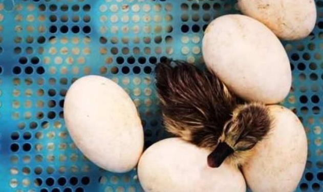


Incubation period of duck eggs
The incubation period of poultry refers to the time from the hatching of eggs to the hatching of chicks under normal economic conditions. The incubation period of different locust poultry species and different production uses is also slightly different: the incubation period of meat duck eggs is slightly longer.
Generally speaking, the incubation period of duck eggs is 28 days, and that of rumen ducks is 33-35 days. The incubation period is shortened when the incubation temperature is higher during the incubation process; When the temperature of hepatization is low, the incubation period is prolonged and the hatching is delayed. Whether the hatching time is delayed or advanced, it is an abnormal performance of hatching. There are more weak ducklings and the survival rate of ducklings is also low.
2、 Embryo growth and development
The embryonic development of poultry is divided into the following two stages:
(1) The development of the embryo during egg formation, after the yolk is discharged from the ovary; Funnel of fallopian tube
to accept; They meet with sperm to fertilize, become a fertilized egg, and begin to develop during egg formation.
When the fertilized egg reaches the isthmus, it will split and reach 256 cell stages 4-5 hours after entering the uterus. When the egg is produced, the embryo development has entered the blastocyst stage or the early gastrula stage. After the egg is produced, the temperature outside the day is lower than the temperature required for the embryo development, and the embryo development is at a standstill. With the extension of time, the embryo gradually dies, which is the reason why the egg preservation time is too long to reduce the incubation rate.
(2) The development of the embryo in the incubation period, when the appropriate incubation conditions are given to the seed egg, the embryo from dormancy
Wake up and continue to develop into chicks. The development of the embryo at different days of age is summarized as follows: on the first day, the embryo defense undergoes primitive metabolism by osmosis, and the primordium of organs such as the archeline, chordal process and vascular region appears, and the dark area of the embryo disc is significantly expanded.
The diameter of the liver disc reached 1.6-2.4 mm when the primary line was born; The bright area is 1.2-1.6 mm, and the dark area is 0.5-0.6 mm wide. When illuminating the egg, the embryo disc becomes a slightly bright dot, commonly known as "white light bead".
The next day, the blastoderm enlarged. The notochord expanded to form five brain bubbles, the brain and notochord began to form neural tubes, and the eye bubbles protruded outward. The heart forms and begins to beat, and the yolk blood circulation begins. The diameter of duck embryo reaches 2.5-2.7 mm, and the length of vascular region is 4.5-7 mm. When you look at the egg, you can see that the dot is more than that of the previous day, commonly known as "fish eye".
On the third day, the vascular area was round, the head was obviously bent to the left and perpendicular to the body, and the amniotic membrane developed to the position of the yolk artery. There are three pairs of cheek cracks and tail buds. The diameter of the embryo is 5.0-6.6 mm, and the transverse diameter of the vascular area is 20-22 mm. When the egg is photographed, the blood vessel area of the embryo looks like cherry.
Before the fourth day, the cerebral vesicles protruded laterally and began to form hemicerebrum. The embryo body was further bent, and the beak, limbs, viscera and allantoic primordium appeared. Due to the obvious expansion of egg yolk due to the infiltration of protein and water, the amniotic cavity forms bifurcated mosquito-like, commonly known as "mosquito beads".
On the fifth day, the embryonic head was significantly enlarged and separated from the egg yolk. The forebrain began to be divided into two hemispheres, and the fifth pair of trigeminal nerves developed. The mouth begins to form, the frontal process grows, and the eyes have obvious pigmentation. Spleen and germ cells lay the foundation. The blood vessels of the egg sac are attached to the egg shell, which is easy to exchange gas through the pores of the egg shell, thus generating heat energy. The allantoic sac rapidly enlarged to form a stalked sac with a diameter of 5.5-6 mm. When the egg is photographed, the yolk blood vessel looks like a little spider, also known as "little spider".